Information For Doctors
Further enhance your practice by offering state of the art technology without the hassle or cost of buying your own CBCT machine!
Just give your patient our referral slip and they can schedule an appointment at their convenience and we will come to them! It's as easy as that! Patients don't have to fight traffic driving to another facility or dread sitting in a waiting room for their name to be called. With our convenient service, they have the comfort of their own home or workplace. Our quick turn around time will provide the results emailed or delivered to your office before the patient's next scheduled appointment.
Referral Form
As with all dental imaging centers, state law requires we have a referral form stating what the patient needs scanned and the doctor's authorization.
After filling out the referral form, please fax or email a copy to our office to ensure that we have it. You may then give it to the patient so that they can call us to schedule an appointment at their convenience or put their name and contact info on it and we will call them to schedule the appointment once faxed or emailed.
Please let the patient know that there is a fee for this scan unless you would like us to bill your office directly. If the patient is taking care of the payment with us, we accept all forms of payment including Visa, MasterCard, Discover and American Express. Unfortunately we do not accept CareCredit. Reimbursement paperwork for their insurance provider will be provided to the patient when payment is collected.
Surgical Implant Guides
Guided implant placement is a technology for accurately placing implants in the jawbone for optimal function and aesthetic restoration.
Using CBCT and 3D Imaging, dentists can now visualize the placement of dental implants in 3 dimensions. This eliminates the guesswork involved in determining what parts of the jawbone offer the best site for dental implant placement.
To carry out the surgery as planned, a custom surgical template is made and used with a guided implant surgery kit.
DICOM files can be provided for Guided Surgery at any time. Below are some of the Guided Surgery companies that we work with regularly.
Haupt Dental Lab
(714) 529-9792
Implant Concierge
(866) 977-2228
Spectrum Dental Lab
(800) 778-4196
Si-Guide Dental Lab
855-373-1614
Results
Results can be printed on photographic quality paper at a 1:1 ratio of
the patient.
The images are available through email or CD in a JPEG or PDF format. Click below to download Adobe to your computer to view PDF formats.
We can provide a free Interactive software of your patient's scan upon request. A tutorial for navigating the software is available any time.
Our CBCT scans can be converted to DICOM files to be viewed in 3rd party viewing software. Click below to download a free DICOM viewing software.

Let US help bring efficiency to your PRACTICE.
Implant Workup
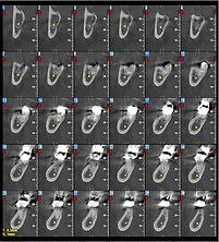
Study consists of panoramic projections and 1 mm cross sections. IAN Nerve in the mandible will be localized and marked. Measurements will be provided from the crest of the bone to the sinus or nerve as well as buccal/lingual width measurements where patient is missing teeth or on compromised teeth that are requested. This workup can be of an individual area or the entire maxilla and/or mandible. 3D rendered images are also provided.


TMJ Workup
Study consists of the lateral and anterior temperal-mandibular joint. This study can be done both in the closed and open positions. Additional positions such as at rest and with/without a splint can also be requested. Mesial/Distal cross sections through both condyles will be provided in each position for visualization of the condyle placement in the joint. Transaxial views and 3D rendering are included as well.


Impaction/Localization Study
Study consists of specific area of interest. Sagittal, coronal and axial cross sections are provided to better visualize tooth of interest location to surrounding structures. 3D images of teeth are included for visualization.


Tissue Pathology
Study consists of panoramic projections along with sagittal, coronal and axial cross sections of the area of interest.


Sinus Evaluation
Study consists of panoramic projections of the maxillary sinus along with sagittal, coronal and axial views of the entire maxillary sinus up to the orbits.


3rd Molar Extraction Study
The IAN nerve will be mapped in the mandibular arch for nerve/root relationship. Mesial/distal and buccal/lingual cross sections are provided to show
proximity to sinus/nerve.


Panorex
Full Mouth Visualization

Radiology Report
If you need Radiographic Interpretation of areas of interest or captured anatomy around the requested area of interest then we can have the scan read by highly experienced Maxillo-Facial Radiologists.
Radiology Reports are not included with our scans but are recommended to be ordered. A radiology report can be requested for an additional fee and will be sent to UCLA to be read. Please allow a few days to receive the report or let us know if a RUSH is needed.
_PNG.png)